
Bacterial Screening of Platelets: Why Culture Media Design Matters
Written By: AnalytiChem |
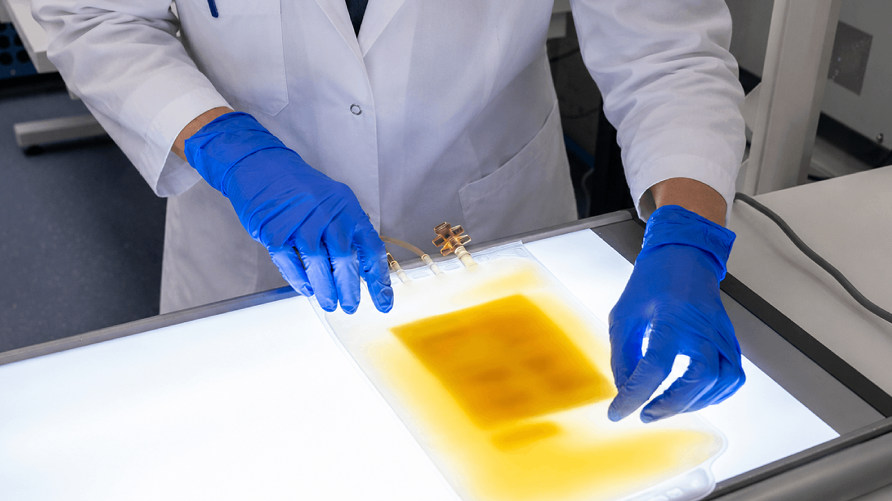
How sampling, timing and culture media design determine what a negative platelet screen actually means.
In this article, you’ll learn:
-
Why platelets carry a bacterial contamination risk that other blood components don’t
-
How contamination rates are reported differently across CDC, FDA, and systematic-review estimates
-
How the UK, EU, and USA each require bacterial risk control for platelets
-
Why a negative bacterial culture doesn’t prove a platelet unit is sterile
-
How aerobic and anaerobic culture media formulation affects detection
- Why false negatives still occur, and what this means for culture media specification
A negative bacterial culture is not proof that a platelet unit is sterile. It means only that no growth reached the detection threshold in the volume that was tested, which is a narrower statement than it first appears. Because platelets can only be stored under conditions that also favor bacterial growth, how reliably that sample is drawn and tested has a direct bearing on patient safety.
In this context, bacterial screening means the culture-based and other validated methods used to detect bacterial contamination in a platelet component before it’s released for transfusion.
Among transfusable blood components, platelets stored at room temperature present a distinctive microbiological challenge. Storage at approximately 20-24 °C preserves platelet function, but also permits contaminating bacteria to multiply. That single physical constraint is why platelets, alone among routine components, attract a dedicated bacterial control requirement in every major regulatory framework.
The size of the residual risk depends heavily on how it’s measured. U.S. Centers for Disease Control and Prevention (CDC) clinical guidance cites contamination of approximately one in 2,000 to 2,500 platelet units[1], and the US Food and Drug Administration (FDA) has estimated residual bacterial contamination at roughly one in 2,500 transfused units[2]. Reported septic-reaction rates range from around one in 100,000 under passive surveillance to one in 10,000 under active surveillance[2], which indicates as much about surveillance design as about the underlying hazard. A 2020 systematic review of twenty-two primary-culture studies estimated an overall culture-positive contamination rate of 0.51 per 1,000 components — approximately one in 1,961 — and found that the measured rate varied with collection method, sample volume, positivity criteria, and study period[3].
These figures are not interchangeable. They describe different populations, different detection strategies, and different endpoints. Reading them as a single number is the first analytical error in this field.
The organisms are consistent across regions. Skin flora, particularly coagulase-negative staphylococci such as Staphylococcus epidermidis, are recovered most frequently. Gram-negative organisms, including Serratia, Enterobacter, and Salmonella species, are encountered less often, but are associated with more severe and sometimes fatal infections[1]. Staphylococcus aureus fits neither description neatly. It’s skin flora, acquired by the same route as the coagulase-negative staphylococci, but is responsible for a disproportionate share of serious outcomes, and is the organism most often recovered when a component fails despite a negative screen. More recent investigations have also implicated Acinetobacter calcoaceticus–baumannii complex, Staphylococcus saprophyticus, and Leclercia adecarboxylata, including polymicrobial contamination traced to platelet-collection-set manufacturing[4].
Three frameworks, one objective
UK
UK blood establishments and hospital blood banks operate under the Blood Safety and Quality Regulations 2005, as amended. The legislation requires authorization, quality systems, traceability, reporting of serious adverse reactions and events, and appropriate bacteriological control of collection and manufacture[5]. The Medicines and Healthcare products Regulatory Agency (MHRA) is the competent authority. Blood establishments require authorization, are inspected at least every two years, and must report serious events and reactions through MHRA’s online reporting system, Serious Adverse Blood Reactions and Events (SABRE)[6][7]
Operational expectations sit one level below the statute, in the Joint United Kingdom (UK) Blood Transfusion and Tissue Transplantation Services Professional Advisory Committee (JPAC) Guidelines for the Blood Transfusion Services in the UK, known as the Red Book. JPAC is not itself legislation, but its specifications are the authoritative common service standard for the UK Blood Services. It requires a validated means of detecting bacterial contamination in platelet components, with an effective sample size, either rapid detection or continuous automated monitoring, and reliable detection at levels relevant to recipient risk[8].
The outcome data are particularly compelling. In the original NHS Blood and Transplant (NHSBT) evaluation, platelet components were sampled from 36 hours after donation, with 8 ml inoculated into each of an aerobic and an anaerobic culture bottle. Among more than 1.2 million components screened over 2011–2015, the confirmed bacterial positivity was 0.03%. Four false-negative cultures were identified, all involving S.aureus. Three were detected by visual inspection before transfusion, while one resulted in recipient morbidity. Compared with the five-year period immediately before implementation of bacterial screening (2006–2010), clinically adverse bacterial transmissions decreased by approximately 90%[9].
Updated English data covering 2014–2023 included more than 2.7 million platelet donations and identified bacterial contamination involving seventy-eight species. Confirmed contamination was more frequent in pooled than in apheresis platelets, at 0.09% versus 0.02%. S. aureus accounted for one confirmed bacterial transmission and eight of ten bacterial near misses[10]. Delayed, large-volume culture markedly reduces risk without making the system infallible.
EU
Regulation (EU) 2024/1938 on substances of human origin came into force on 7 August 2024 but, with limited exceptions, applies from 7 August 2027, when it repeals Directives 2002/98/EC and 2004/23/EC. It requires risk-based quality systems, validated processing and equipment, microbial contamination controls, documented release, vigilance, and competent-authority oversight. It also permits Member States to retain more stringent national measures, which means convergence on principles rather than uniformity of protocol[11][12].
That distinction matters for anyone assuming the Regulation will impose a single European platelet-culture method — this is not the case. Operational bacterial control remains strongly dependent on European Directorate for the Quality of Medicines & HealthCare (EDQM) guidance, national competent authorities, and validated blood-establishment procedures[13]. The European Centre for Disease Prevention and Control (ECDC) is developing communicable-disease guidance for substances of human origin, but its published program as of 2026 primarily addresses donor-transmitted pathogens such as HIV and hepatitis viruses, and should not be cited as though it currently prescribes a pan-European bacterial screening protocol[14].
European vigilance data reflects this heterogeneity. The European Commission’s compilation of 2023 national reporting recorded forty-one transfusion-transmitted infection cases, of which thirty were bacterial. Five were fatal, and four of those five followed platelet transfusion[15]. Cross-country interpretation requires caution because reporting completeness, case definitions, and denominator quality differ between Member States.
France offers the most instructive organism-level detail. Its 2024 hemovigilance report describes a serious infection involving pathogen-reduced apheresis platelets and Staphylococcus ureilyticus, with an unfavorable outcome[16]. The published case series records that the patient died within hours of transfusion, and also identified Bacillus mobilis contamination in a pathogen-reduced pooled component. Proposed mechanisms for residual risk after pathogen reduction include spores, biofilms, high starting bioburden, contamination occurring after treatment, and organisms with reduced susceptibility to the treatment process[17].
The surveillance lesson is symmetrical. Culture is vulnerable to sampling error and to slow or spatially uneven growth. Pathogen reduction is vulnerable to biological resistance, process limitations, and post-treatment contamination. Neither removes the need for visual inspection, traceability, prompt clinical recognition, and investigation.
USA
The binding foundation is 21 CFR 606.145, which requires blood collection establishments and transfusion services to assure that the risk of bacterial contamination of platelets is adequately controlled using FDA-approved or -cleared devices, or other adequate and appropriate methods found acceptable by the FDA. Identified contaminated units and associated components must not be released, and organism identification and notification actions are required[18].
The FDA’s December 2020 guidance then sets out acceptable risk-control strategies, including large-volume delayed sampling, primary culture followed by secondary culture or rapid testing, and pathogen reduction. Seven-day dating requires appropriately cleared or approved storage containers together with bacterial-detection devices labeled for the applicable safety-measure use[2]. Association for the Advancement of Blood & Biotherapies (AABB) Association Bulletin #21-02, updated in March 2026, translates these options into implementation considerations, while stressing that no single strategy eliminates every contaminant[19].
The USA’s framework is therefore the most procedurally explicit of the three. It specifies sampling times and volumes directly, where the UK works through service-wide specifications, and the EU works through risk-based obligations delegated to national authorities. All three arrive at the same operational conclusion: sample late enough, sample sufficient volume, use a validated detection system, and keep inspecting.
A negative culture is not proof of sterility
Culture screening examines a sample, not every organism in the platelet bag. If contamination is present in only a few cells per unit, an early or small-volume sample might contain no organisms at all. This is a Poisson-limited sampling problem. An excellent culture medium can’t recover a bacterium that never enters the bottle.
Two levers address this. Delayed sampling gives viable contaminants additional time to multiply, and increased sample volume raises the probability that at least one cell is captured. That’s why large-volume delayed sampling represents more than a procedural adjustment.
The FDA’s single-step Large Volume Delayed Sampling (LVDS) strategy for five-day platelets calls for a single culture sampled no sooner than thirty-six hours after collection, using a sampling volume of at least 16 ml inoculated evenly into aerobic and anaerobic culture media, with a minimum incubation period of twelve hours before release. For appropriately labeled apheresis-platelet systems supporting storage through day seven, the corresponding single-step sample is taken no sooner than forty-eight hours[2].
Culture sensitivity is therefore an end-to-end property of the workflow. Contamination level, organism physiology, sampling time, sampled volume, distribution within the bag, culture atmosphere, media formulation, incubation, and instrument algorithm all contribute. Change one, and the validated performance of the whole is changed.
“The question I would want a blood service to ask is not whether a medium grows the organism, but whether it does so reproducibly in a platelet matrix, in the atmosphere it will actually be used in, and within the time the component is still in date. Growth promotion on a reference strain answers a much easier question than the one the screening workflow is really asking.”
— Yoggya De Silva, Product Manager, Culture Media, Life Sciences Portfolio, AnalytiChem Group
What happens after an organism reaches the bottle?
Automated platelet-culture systems generally infer growth from microbial metabolism. FDA-cleared systems described in public 510(k) premarket notification summaries, FDA submissions used to demonstrate substantial equivalence to an existing legally marketed device, incubate inoculated bottles and detect increasing carbon dioxide through a colorimetric or fluorescent sensor. The limitation is explicit in the labeling: detection is restricted to viable organisms that can grow in the particular medium and generate a detectable metabolic signal[20].
As a result, platelet-culture media products are not generic laboratory broths. An FDA submission for one cleared system describes an aerobic formulation based on casein and soybean digests, sodium polyanethol sulfonate, pyridoxine, and complex nutrient substrates in an oxygen-containing atmosphere. Its paired anaerobic bottle additionally contains yeast extract, hemin, menadione, pyruvate, and reducing agents in a nitrogen-containing atmosphere. The two bottles create different nutritional and atmospheric recovery conditions, and are cleared for different organism groups[20].
Clinical evidence supports the pairing. In experiments with a Staphylococcus lugdunensis isolate implicated in a fatal platelet transfusion, larger inoculated volumes improved recovery at low contamination levels, and when only one or two organisms entered a bottle, the anaerobic medium became reactive sooner than the aerobic bottle[2]. At that scale, hours of time to detection are potentially the difference between interdiction and transfusion.
The incremental benefit is small in absolute terms but nonetheless real, and carries a cost. A systematic review covering more than 1.7 million platelet components found that, after excluding duplicate aerobic detections and Cutibacterium or Propionibacterium, anaerobic culture uniquely detected approximately 0.06 contamination events per 1,000 components, around one in 17,000 tested[21]. Those genera are excluded because they do not proliferate in the oxygenated storage environment, and so are not thought to reach levels capable of causing an acute transfusion reaction [22]. A separate meta-analysis estimated false-positive rates of 1.0 per 1,000 for aerobic culture, 1.8 per 1,000 for anaerobic culture, and 2.4 per 1,000 when both were used[23]. Broader recovery conditions and higher discard rates are two faces of the same decision.
Fastidious organisms expose formulation limits
Cutibacterium acnes illustrates why growth promotion must be assessed against the organism, the matrix, and the time to detection, rather than against eventual growth. It’s an aerotolerant anaerobe, so survives in the oxygenated component without multiplying, and only grows once it reaches the anaerobic bottle. That’s why it’s excluded from acute-risk estimates, and why its significance is unsettled rather than dismissed: it doesn’t reach levels associated with acute septic reactions, but is a recognized cause of endocarditis and prosthetic infection, and has been proposed as a cause of unrecognized chronic transfusion-transmitted infection [22]. Reported mean times to detection in platelet components sit at around three days, with a substantial tail extending well beyond five days, which means affected platelets can be issued before the bottle signals [24].
In a 2023 proof-of-principle study, supplementation of anaerobic culture bottles with a commercial vitamin and cofactor mixture accelerated the detection of C. acnes in platelet components, with the largest observed reduction in time to detection being approximately 1.2 days relative to unsupplemented bottles. Tween 80 produced even greater growth enhancement in broth, but platelet- and plasma-containing bottles generated false-positive instrument signals[25]. The study demonstrates both sides of media design: additional nutrients may accelerate a difficult organism, yet a formulation change can interact unexpectedly with the sample matrix or the sensor.
This isn’t an argument for modifying cleared bottles in routine practice, and the regulations foreclose it in any case. The FDA requires bacterial testing to be performed consistently with the device instructions for use, and 21 CFR 606.65(e) requires reagents and supplies to be used according to manufacturer instructions[26][2]. The study is evidence for manufacturers developing future formulations. Faster growth is valuable only when specificity, matrix compatibility, and system performance remain controlled.
Why do false negatives still occur?
False-negative platelet cultures arise through several mechanisms, which are not mutually exclusive:
- Sampling error: where very low bacterial numbers are simply absent from the aliquot removed from the bag.
- Sampling too early: when the organism remains below a reliably sampled concentration or is still in lag phase.
- Insufficient sample volume: which transfers fewer organisms and reduces recovery probability.
- Atmospheric mismatch: where an obligate anaerobe is missed by an aerobic-only strategy, or a facultative organism signals sooner in an anaerobic formulation.
- Slow or fastidious growth: where the organism reaches the instrument threshold only after the component has been released or transfused.
- Biofilm formation or non-uniform distribution: Serratia marcescens isolates capable of forming platelet-associated biofilms were roughly three times more likely to be missed by automated culture than biofilm-negative strains in one study[27].
- Workflow or device deviations: including reduced inoculation volume, incompatible media, altered incubation, or use outside the cleared indication.
These mechanisms are not evenly distributed across organisms. A platelet concentrate is not a passive broth: platelets sense bacteria and release antimicrobial factors including platelet factor 4 and thrombocidins, so the organisms that persist are those able to evade them. S. aureus is well equipped to do so, through surface modification, capsule formation, and degradation of antimicrobial peptides. Its occasional escape from screening is attributed principally to delayed growth and biofilm formation during storage [28]
Clinical surveillance confirms that a negative primary culture doesn’t eliminate risk. In one seven-year active-surveillance study, twenty of more than 51,000 transfused platelet units were bacterially contaminated despite having been cultured after collection and released as negative. Five recipients developed septic transfusion reactions, none of which were captured by passive surveillance[29].
UK experience in 2025 made the same point from the other direction. The NHSBT investigated one hundred suspected bacterial incidents that year. No bacterial transmission was confirmed, but three near misses involved S. aureus: one platelet pool developed a white deposit, and two components developed aggregates, all despite negative routine screening, and retesting recovered the organism. Over 1996–2025, Serious Hazards of Transfusion (SHOT) recorded thirty-seven confirmed bacterial incidents affecting forty recipients, thirty-three of whom had received platelet components. The most recent confirmed bacterial transmission was reported in 2015[30].
These UK haemovigilance findings illustrate an important limitation of culture-based screening and help clarify what a “negative-to-date” result actually means. It indicates that bacterial growth has not been detected in the inoculated sample at that point in the monitoring period. This may be because organisms are absent from the sampled aliquot or because any organisms present have not yet multiplied sufficiently to trigger the detection system. Therefore, a negative-to-date result should not be interpreted as evidence that the entire platelet component is sterile.
What does this mean for media specification?
From the perspective of a manufacturer of ready-made culture media, the defensible claim isn’t that better broth prevents all false negatives. Media can’t correct an unrepresentative sample, an unsuitable sampling time, biofilm sequestration, or use outside a validated system.
What well-designed ready-made media can contribute is a controlled and documented growth environment within the performance envelope of the complete detection system. A meaningful quality package should show how formulation, fill volume, atmosphere, container, sensor compatibility, shelf life, and organism recovery are each controlled against established specifications, and how lot-to-lot consistency is demonstrated.
For a new bacterial-detection method, the FDA recommends a validation plan and a summary of validation data. Public 510(k) evidence offers a useful, though not universally prescriptive, example: one platelet-culture system was evaluated at two sites across three media lots, using platelet matrices seeded at a target of approximately 1 CFU/ml, with organism-specific time-to-detection assessment and terminal subculture of instrument-negative bottles [2] [20]. The underlying quality expectation has also tightened. For device manufacturers in the USA, the FDA Quality Management System Regulation has incorporated ISO 13485:2016 since 2 February 2026[31].
Platelet screening sensitivity is designed into the full method. Delayed and sufficiently large sampling improves the chance that contamination reaches the bottle. Complementary aerobic and anaerobic media broaden recovery conditions. Carefully validated nutrients and atmospheres support clinically relevant organisms. Consistent lot performance ensures the validated method is reproduced in routine use. None of these is sufficient alone, but together they form a more reliable barrier against transfusion-transmitted bacterial infection.
Room-temperature storage is not a convenience; it’s the condition conventional platelets need to retain their hemostatic function, and is the same condition that allows contaminating bacteria to survive and multiply. Sampling strategy, culture media formulation, and regulation each manage that constraint from a different angle, and the surveillance data from the UK, EU, and USA show that this combined approach, not any single safeguard, has reduced bacterial transmission.
Redipor® prepared media products are manufactured by AnalytiChem for microbiological quality control applications, with formulation, fill, and lot performance controlled to documented specifications. For questions about media specification in a validated screening workflow, contact our technical team.
Frequently Asked Questions
Can pathogen reduction replace bacterial culture screening for platelets?
Not on current evidence. France’s 2024 hemovigilance report describes a serious infection involving pathogen-reduced apheresis platelets and Staphylococcus ureilyticus, with an unfavorable outcome; in the published case series, the patient died within hours of transfusion, and Bacillus mobilis contamination was identified in a pathogen-reduced pooled component. Proposed mechanisms for this residual risk include spores, biofilms, high starting bioburden, contamination occurring after treatment, and organisms with reduced susceptibility to the treatment process, so pathogen reduction is vulnerable in ways that culture is not, and neither method removes the need for visual inspection and investigation.
Why are pooled platelets contaminated more often than apheresis platelets?
Does visual inspection catch contamination that culture screening misses?
Is donor skin flora responsible for all platelet bacterial contamination?
Subscribe to our blog
References
1. CDC. Blood safety: clinical guidance for healthcare providers. https://www.cdc.gov/blood-safety/hcp/clinical-guidance/index.htmL
2. FDA. Bacterial Risk Control Strategies for Blood Collection Establishments and Transfusion Services to Enhance the Safety and Availability of Platelets for Transfusion: Guidance for Industry. December 2020. https://www.fda.gov/media/123448/download
3. White SK, et al. Bacterial contamination rate of platelet components by primary culture: a systematic review and meta-analysis. Transfusion. 2020. https://pubmed.ncbi.nlm.nih.gov/32181889/
4. Kracalik I, Kent AG, Villa CH, et al. Posttransfusion sepsis attributable to bacterial contamination in platelet collection set manufacturing facility, United States. Emerg Infect Dis. 2023;29(10):1979-1989. https://doi.org/10.3201/eid2910.230869
5. The Blood Safety and Quality Regulations 2005 (SI 2005/50), as amended. https://www.legislation.gov.uk/uksi/2005/50/contents
6. MHRA. Blood regulation and safety. GOV.UK. https://www.gov.uk/government/collections/blood-regulation-and-safety
7. MHRA. Blood: authorisations and safety reporting (SABRE). GOV.UK. https://www.gov.uk/guidance/blood-authorisations-and-safety-reporting
8. JPAC. Guidelines for the Blood Transfusion Services in the UK, section 9.5: Recommended standards for the reduction of bacterial contamination of blood components. https://www.transfusionguidelines.org/red-book/chapter-9-microbiology-tests-for-donors-and-donations-general-specifications-for-laboratory-test-procedures/9-5-recommended-standards-for-the-reduction-of-bacterial-contamination-of-blood-components
9. McDonald C, et al. Bacterial screening of platelet components by National Health Service Blood and Transplant, an effective risk reduction measure. Transfusion. 2017. https://pubmed.ncbi.nlm.nih.gov/28425610/
10. Chugh V, Secret S, Davison K, St John A, Simmonds P, Brailsford SR, Harvala H. Bacterial screening of platelet donations in England, 2014-2023. Vox Sanguinis. 2026. https://pmc.ncbi.nlm.nih.gov/articles/PMC13356991/
11. Regulation (EU) 2024/1938 on standards of quality and safety for substances of human origin intended for human application. https://eur-lex.europa.eu/eli/reg/2024/1938/oj
12. European Commission. SoHO Regulation: new EU rules on substances of human origin. https://health.ec.europa.eu/blood-tissues-cells-and-organs/soho-regulation_en
13. EDQM. Guide to the preparation, use and quality assurance of blood components, 22nd edition. https://www.edqm.eu/en/blood-guide
14. ECDC. Technical guidelines: substances of human origin. https://www.ecdc.europa.eu/en/infectious-disease-topics/related-public-health-topics/substances-human-origin/technical-guidelines
15. European Commission. Summary of the 2024 (2023 data) annual reporting of serious adverse reactions and events for blood and blood components. https://health.ec.europa.eu/latest-updates/summary-2024-2023-data-annual-reporting-serious-adverse-reactions-and-events-blood-and-blood-2025-08-26_en
16. ANSM. French haemovigilance annual report, year 2024: executive summary. https://ansm.sante.fr/uploads/2025/10/23/20251023-hemovigilance-rapport-2024-synthese-en.pdf
17. Laperche S, et al. Pathogen-reduced platelet concentrates in France: impact on the risk of transfusion-transmitted infections, 2017-2024. Vox Sanguinis. 2026. https://pmc.ncbi.nlm.nih.gov/articles/PMC13158009/
18. 21 CFR 606.145: Control of bacterial contamination of platelets. https://www.ecfr.gov/current/title-21/section-606.145
19. AABB. Association Bulletin #21-02: Considerations for Implementation of FDA Guidance on Bacterial Risk Controls for Platelets. Updated March 2026. https://www.aabb.org/docs/default-source/default-document-library/resources/association-bulletins/ab21-02-revised.pdf?sfvrsn=21ff9c05_3
20. FDA. 510(k) Premarket Notification database (searchable summaries for cleared bacterial-detection systems). https://www.accessdata.fda.gov/scripts/cdrh/cfdocs/cfpmn/pmn.cfm
21. Corean J, et al. The incremental benefit of anaerobic culture for controlling bacterial risk in platelets: a systematic review and meta-analysis. Vox Sanguinis. 2021. https://onlinelibrary.wiley.com/doi/10.1111/vox.13013
22. Ramirez-Arcos S, McDonald C. Bacterial contamination of platelet components. Annals of Blood. 2022;7:24. doi:10.21037/aob-21-84. https://aob.amegroups.org/article/view/6846/htmL
23. Corean J, et al. Platelet component false positive detection rate in aerobic and anaerobic primary culture: a systematic review and meta-analysis. Transfusion Medicine Reviews. 2021;35(3). https://pubmed.ncbi.nlm.nih.gov/34158212/
24. Daane L, Adamik M, Ullery M, Ahmed M, Yang J. Rapid detection of bacterial contaminants in platelet concentrates using the next generation BACT/ALERT VIRTUO Microbial Detection System. Annals of Blood. 2021;6. https://aob.amegroups.org/article/view/6468/htmL
25. Kumaran D, et al. Nutrient supplementation of culture media improves the detection of Cutibacterium acnes in platelet components by an automated culture system. Vox Sanguinis. 2023. https://onlinelibrary.wiley.com/doi/10.1111/vox.13529
26. 21 CFR 606.65: Supplies and reagents. https://www.ecfr.gov/current/title-21/section-606.65
27. Greco-Stewart VS, et al. Serratia marcescens strains implicated in adverse transfusion reactions form biofilms in platelet concentrates and demonstrate reduced detection by automated culture. Vox Sanguinis. 2012. https://pubmed.ncbi.nlm.nih.gov/21967170/
28. Chi C, Ramirez-Arcos S. Platelet concentrates safety: a focus on the challenging pathogen Staphylococcus aureus, a narrative review. Annals of Blood. 2025;10:5. doi:10.21037/aob-24-31. https://aob.amegroups.org/article/view/10639/htmL
29. Hong H, et al. Detection of septic transfusion reactions to platelet transfusions by active and passive surveillance. Blood. 2016;127(4):496. https://ashpublications.org/blood/article/127/4/496/35018/
30. Serious Hazards of Transfusion. Annual SHOT Report 2025, chapter 24: Transfusion-Transmitted Infections. https://www.shotuk.org/shot-reports/annual-shot-report-2025/
31. FDA. Quality Management System Regulation (QMSR). https://www.fda.gov/medical-devices/postmarket-requirements-devices/quality-management-system-regulation-qmsr